
Have you ever wondered why there are different types of imaging tests? Or what the differences between the many types of imaging tests are? Learn about the possible modalities and their purposes here.
Different Types of Imaging Tests: Sorting Out the Differences
If your doctor has ordered a medical imaging exam for you, you might have questions about the type of scan or test you will be having. Technologists use modalities (different types) to gather the right images for your radiologist to examine. If you’re getting a scan to see if you have a concussion, for instance, CT would be the modality for your exam. On the other hand, if you are getting a mammogram, X-ray would be the modality in use.
At UVA Radiology and Medical Imaging, we use each modality to perform multiple types of imaging tests to diagnose multiple kinds of conditions. Each modality is unique in terms of the images it gathers, equipment it uses, and conditions it helps radiologists diagnose. Learn more about our five most common modalities for our various types of imaging tests: X-ray, CT, MRI, ultrasound, and PET.
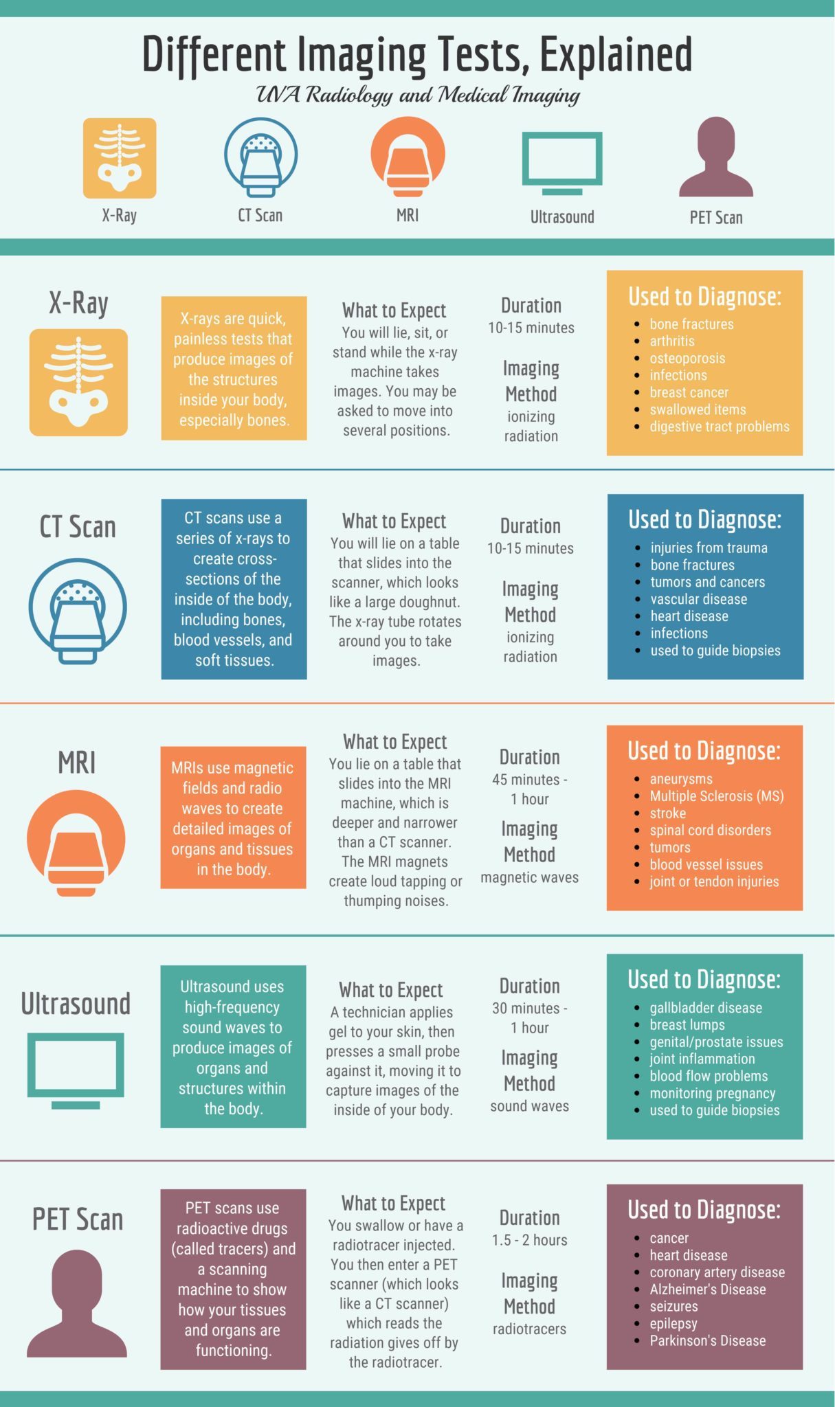
Infographic Text: Different Imaging Tests, Explained
UVA Radiology and Medical Imaging
X-Ray, CT Scan, MRI, Ultrasound, PET Scan
X-Ray:
-
X-rays are quick, painless tests that produce images of the structures inside your body, especially bones.
-
What to expect: You will lie, sit, or stand while the x-ray machine takes images. You may be asked to move into several positions.
-
Duration: 10-15 minutes
- Imaging Method: ionizing radiation
-
Used to diagnose: bone fractures; arthritis; osteoporosis; infections; breast cancer; swallowed items; digestive tract problems
CT Scan:
-
CT scans use a series of x-rays to create cross-sections of the inside of the body, including bones, blood vessels, and soft tissues.
-
What to expect: You will lie on a table that slides into the scanner, which looks like a large doughnut. The x-ray tube rotates around you to take images.
-
Duration: 10-15 minutes
- Imaging Method: ionizing radiation
-
Used to diagnose: injuries from trauma; bone fractures; tumors and cancers; vascular disease; heart disease; infections; used to guide biopsies
MRI:
-
MRIs use magnetic fields and radio waves to create detailed images of organs and tissues in the body.
-
What to expect: You lie on a table that slides into the MRI machine, which is deeper and narrower than a CT scanner. The MRI magnets create loud tapping or thumping noises.
-
Duration: 45 minutes – 1 hour
- Imaging Method: magnetic waves
-
Used to diagnose: aneurysms; Multiple Sclerosis (MS); stoke; spinal cord disorders; tumors; blood vessel issues; joint or tendon injuries
Ultrasound:
-
Ultrasound uses high-frequency sound waves to produce images of organs and structures within the body.
-
What to expect: A technician applies gel to your skin, then presses a small probe against it, moving it to capture images of the inside of your body.
-
Duration: 30 minutes – 1 hour
- Imaging Method: sound waves
-
Used to diagnose: gallbladder disease; breast lumps; genital/prostate issues; joint inflammation; blood flow problems; monitoring pregnancy; used to guide biopsies
PET Scan:
-
PET scans use radioactive drugs (called tracers) and a scanning machine to show how your tissues and organs are functioning.
-
What to expect: You swallow or have a radiotracer injected. You then enter a PET scanner (which looks like a CT scanner) which reads the radiation given off by the radiotracer.
-
Duration: 1.5 – 2 hours
- Imaging Method: radiotracers
-
Used to diagnose: cancer; heart disease; coronary artery disease; Alzheimer’s Disease; seizures; epilepsy; Parkinson’s Disease