
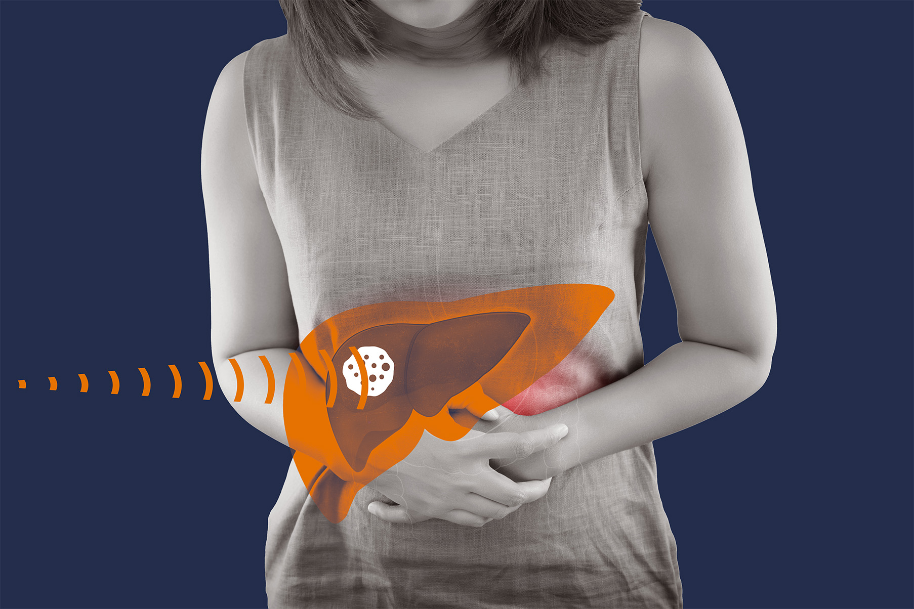
Hepatocellular Carcinoma (HCC) is one of the most common liver cancers and is often caused by long-term damage or scarring of the liver (liver cirrhosis). It is estimated to be responsible for 500,000 deaths every year. For various reasons, including the overall health of the patient, sometimes HCC cannot be treated with surgery. When HCC is inoperable, it continues to cause symptoms and progress as a disease. At UVA Interventional Radiology, we want you to know about radioembolization—a minimally invasive procedure that can offer hope to people suffering from inoperable liver cancer.
What is Radioembolization?
If you have inoperable liver cancer, you do have other options. Radioembolization (also called Y90) is an interventional radiology procedure designed to treat HCC and other inoperable liver cancers. Radioembolization is a palliative procedure, which means it doesn’t cure the cancer. Rather, the goal of this procedure is to relieve HCC symptoms, limit the cancer’s growth, and extend the life expectancy of patients— sometimes long enough time to receive a liver transplant.
In a radioembolization procedure, the interventional radiologist uses radiation-filled glass or resin beads, called microspheres, to block the blood flow to the cancerous growth while delivering radiation to the tumor. The microspheres are about one-third the width of a human hair and filled with yttrium-90 (Y90), a radioactive material. When the microspheres are released in the blood vessel, they deliver a focused, intense amount of radiation to the tumor with little or no radiation delivered to healthy, surrounding tissues, which also results in fewer side effects than typical radiation therapy.
As an interventional radiology procedure, radioembolization is minimally invasive. Interventional radiologists use medical imaging to see inside the patient, allowing them to guide their movements and release the microspheres in real time.
Before the Procedure
Preparing for radioembolization begins with a consultation with an interventional radiologist. This consultation usually involves having a blood test done to test kidney health and how well your blood responds to clotting. Also, make sure to inform your doctor about any medications and herbal supplements you are taking, as well as any known allergies to anaesthesia and contrast dye used for x-rays.
After your initial consultation and testing, the next step is to have an angiogram done the week before the procedure. An angiogram is a map of the blood vessels in your body, in this case, the ones that are connected to the tumor.

As your procedure date approaches, patients are advised to make plans for transport to and from the procedure. Depending on your health, you may go home the same day as your radioembolization, or you may have to stay in the hospital for one or two days.
Because of the radioactive material used in a radioembolization, you will likely be required to limit contact with others after the procedure, especially women who are pregnant and children. Talk with your doctor about specific guidelines, but most patients will have contact restricted for 3-7 days.
How does Radioembolization work?
At the beginning of the radioembolization procedure you may receive a moderate amount of anesthesia or general anesthesia. After that, the interventional radiologist makes a small incision in the groin. Using fluoroscopy, a continuous x-ray to see inside the patient, the interventional radiologist inserts a catheter into the incision and guides it to the femoral artery, the leg’s largest blood vessel.
Once accessing the artery, the catheter is guided to the blood vessel in the liver connected to the tumor. The interventional radiologist then releases the microspheres where they go to work blocking blood flow and delivering radiation to the tumor. The catheter is then removed and the incision is bandaged–stitches aren’t even necessary.

After the procedure, some possible side effects include mild abdominal pain, loss of appetite, nausea, fatigue, and fever. These feelings are usually mild and will disappear over time. Most patients are able to return to normal activities within 1-2 days after their procedure. The interventional radiologist may also schedule follow-up CT or MRI scans to assess the size of the tumor after the procedure and discuss any further treatment options.
Offering Hope
Radioembolization has been shown to shrink the size of liver cancers and a majority of patients will see improvements in the liver, a longer life expectancy, and a reduction in symptoms. Talk to your doctor about your eligibility for radioembolization or contact UVA today at 434.9249401 to learn more.