

Have you been told that your infant needs an ultrasound to screen for hip dysplasia (DDH)? Are you nervous about what it could mean for your child’s health? At UVA Pediatric Radiology, we want parents to learn the facts about hip dysplasia. We also want to calm any fears you may have by showing you how ultrasound screenings can help detect hip dysplasia early, positively influencing your child’s future.
What You Need to Know About Hip Dysplasia (DDH)
Before understanding the importance of screening, it’s necessary to know what hip dysplasia is and how it could affect your child. Developmental Dysplasia of the Hip (DDH), also known as hip dysplasia, affects 2-3 out of every 1,000 babies born worldwide. This condition is present at birth, and is caused by a shallow socket (acetabulum) in the hip joint.

A healthy hip socket should securely hold the “ball”, formally known as the femoral head of the femur bone. With a shallow hip socket, when the “ball” moves back and forth, it can slip out of place because the head of the femur does not fit firmly into the socket.
Parents and doctors can identify potential DDH cases if the child shows these physical symptoms:
- Uneven leg length
- Uneven fat distribution on the thighs
- Lower mobility or flexibility on one side
- Limping, toe walking, or a waddling gait that affects their ability to walk
Should My Infant Be Screened for DDH?
DDH screening is recommended by all the leading U.S. pediatric and orthopedic physician organizations. According to the International Hip Dysplasia Institute, hip ultrasound is suggested for three groups:
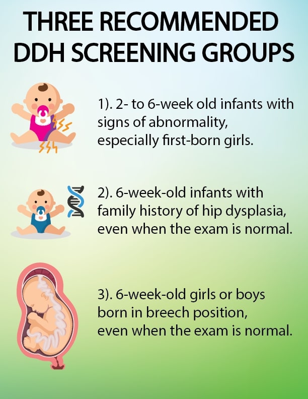
Family history can predispose infants to developing hip dysplasia. You should let your physician know if you or a family member were born with hip dysplasia or have very flexible ligaments. 6 in 10 DDH cases occur in first-born babies and 80% of overall cases occur in females. Even if symptoms do not show during the initial physical examination after birth, it is still important to have your child screened for DDH if your child is in one of these groups. The earlier DDH is diagnosed the more likely treatment will be successful and the more likely less invasive treatments can be used.
DDH is more common in babies born in the breech position. Babies in the normal womb position have less pressure on their hips which is why they are far less likely to have DDH. Hip dysplasia is more common in first-born children because they are often more cramped in the uterus of first-time mothers. There are other less common factors that may increase the chance of an infant having DDH, such as congenital foot and neck anomalies, so a doctor may recommend screening in those cases as well.
What Will My Child and I Experience During a Hip Ultrasound Screening?
At UVA Pediatric Radiology, we offer hip ultrasound screenings to detect the different stages of hip dysplasia before symptoms begin to show. If diagnosed early and treated successfully, children are frequently able to develop a healthy hip joint.
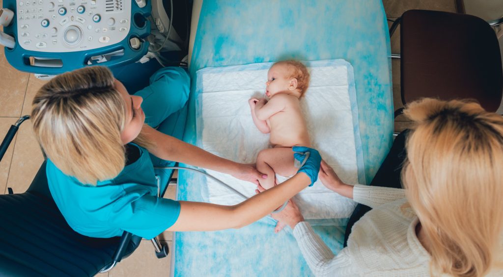
Hip ultrasounds are recommended for infants from birth to six months old because their bones have not fully developed. After about 6 months of age x-rays are done because the bones are often too well developed to use ultrasound successfully. Hip ultrasounds are a safe, non-invasive procedure that does not use any radiation. Ultrasounds use inaudible sound waves which bounce off of the bones and muscles to create an image for radiologists to interpret.
Hip ultrasounds take less than 20 minutes and the child will not feel any pain during the examination. A warm, water-based gel is applied to their skin so the ultrasound can provide better images by taking away air pockets between the transducer and the skin.
Babies must stay as still as they can during the examination, so it is recommended that parents bring toys, or soothe their babies with their voice to make time pass quicker. The technologist will examine both hips of the child in different positions until the images are fully formed. This screening will help doctors to determine if further treatment is needed.
Having Your Child’s Hip Ultrasound at UVA
Hip ultrasound screenings make a major difference in the lives of many children by providing an early diagnosis of hip dysplasia, which can help lead to successful treatment and the development of a healthy hip joint. Children with DDH that go untreated for too long can have life-long problems with walking and pain.
At UVA Pediatric Radiology, we know that children’s bodies are different from those of adults. That’s why we have pediatric radiologists who are specifically trained to provide pediatric radiology services for children of all ages. Additionally, our ultrasound technologists who perform hip ultrasounds complete over 100 hours of technical training to become certified. This sub-specialized training gives UVA an edge in accuracy and efficiency over other medical institutions.
Our goal is to make you and your child’s screening experience as smooth as possible. Talk to your child’s care provider about having your child’s hip ultrasound performed at UVA, or call (434).243.5500 to make an appointment. To learn more about pediatric imaging at UVA, click here.