

Liver disease is a growing issue: by some counts, one-third of the U.S. population has some degree of fatty liver or liver damage. Being able to diagnose fibrosis, or scarring of the liver tissue, as early and accurately as possible is critical, before that damage becomes irreversible and limits your liver’s ability to function.
The standard method of diagnosing fibrosis has long been liver biopsy. But many people are afraid of the pain and the risk of serious complications. A newer, non-invasive technique – elastrography – can be just as accurate as a biopsy for diagnosing fibrosis.
What Does Your Liver Do?
Though it’s not talked about as often as other organs like the heart or the brain, the liver is a big deal – literally! After your skin, it’s the largest organ in your body. And with over 500 different functions, it has a lot of work to do.
The liver’s primary job is filtering everything we eat, drink, or consume. Once food, drink or oral medication is digested by the stomach and intestine, it’s absorbed into our blood. That’s where the liver comes in: the liver filters our blood, removing toxins that can hurt us and storing important nutrients that our body needs to function.
The liver also regulates our blood sugar; produces bile, which breaks down the fat that we eat; removes damaged blood cells from our circulatory system; and plays an important role in our immune response. With all these important jobs to do, it’s critical that doctors know when something is going wrong with your liver.
The Stages of Liver Damage
Liver damage can come from many sources – viruses (like Hepatitis), injury from harmful chemicals, attacks from your own immune system – but no matter the cause, the progression of damage is similar. The four main stages of liver damage are inflammation; fibrosis; cirrhosis; and liver failure.
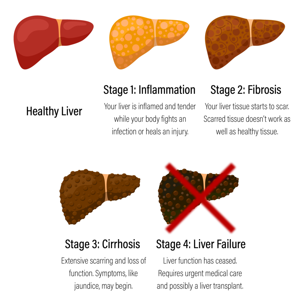
In the inflammation and fibrosis stages, the liver is still able to function normally. And it’s possible for the damage in the liver at these stages to be reversed in some patients. That’s why identifying fibrosis as early and accurately as possible is so important.
How to Determine if You Have Liver Fibrosis
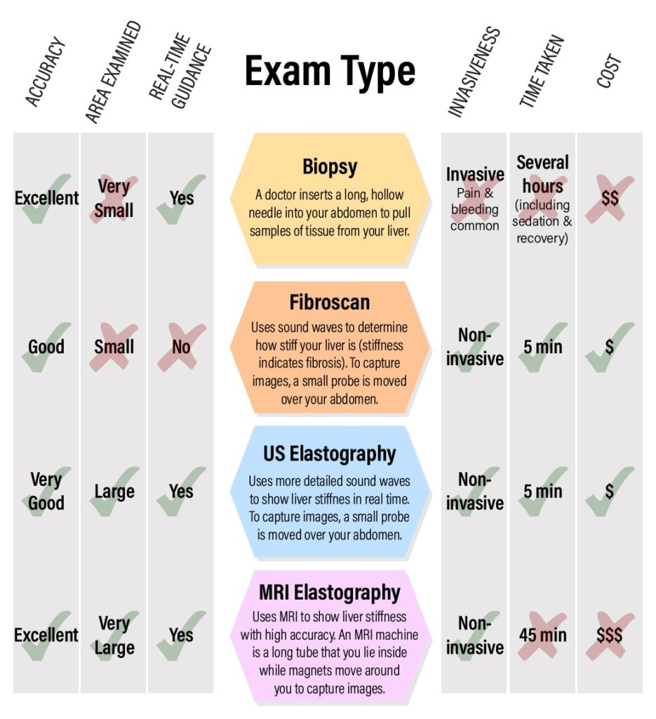
For a long time, liver biopsy has been the standard tool for determining the extent of liver fibrosis. During a biopsy, a doctor inserts a long, thin needle through your abdomen and into your liver. The needle pulls out a small sample of your liver tissue that the doctor can then examine. To capture samples from different parts of your liver, the doctor will have to re-insert the needle 10-15 times for complete analysis.
A biopsy can be painful and lead to complications. And though it’s useful for analyzing the liver tissue for the underlying cause of damage, it can only assess the exact areas where the needle collected tissue. Because of these drawbacks, a number of alternatives have emerged. Some of these alternatives are just as good at identifying fibrosis as a biopsy, and they have the advantage of being painless and non-invasive.
Chart: Comparing Your Options for Identifying Liver Fibrosis
Why Radiologist-Performed Elastrography Might Be Right For You
If you want to avoid a liver biopsy, you might think that the non-invasive options are all the same. But that’s not true: only ultrasound elastography and MRI elastrography are performed by specially trained radiologic technologists and interpreted by radiologists, or doctors who specialize in analyzing medical images.
In addition, only elastography allows doctors to see real-time video of the inside of your liver, which helps them move the probe to exactly where the damage is. Because they can look at the damaged areas in much more detail, your results are more accurate.
Your care provider will determine if ultrasound or MRI elastography is right for you. In general, MRI is recommended only for those cases where more extensive damage is suspected. It’s also an option for those patients with a larger body size for whom ultrasound isn’t as effective.
Elastography at UVA Health
The Department of Radiology and Medical Imaging at UVA Health was one of the first sites in the United States to perform ultrasound elastrography to assess liver fibrosis. We have both the clinical experience and technical ability to perform the exam and analyze the images as quickly and accurately as possible.
At UVA, your ultrasound or MRI technologist is specially trained in obtaining images of the liver using these technologies. We have the latest imaging technology to guarantee the images we obtain are as high-quality as possible. And, most importantly, we have radiologists who are specially trained in analyzing images of the organs in your abdomen. When your health is at stake, you want to choose experts who know exactly what they are looking for.
Talk to your GI doctor about ultrasound or MRI elastography and ask them to refer you to UVA Radiology.