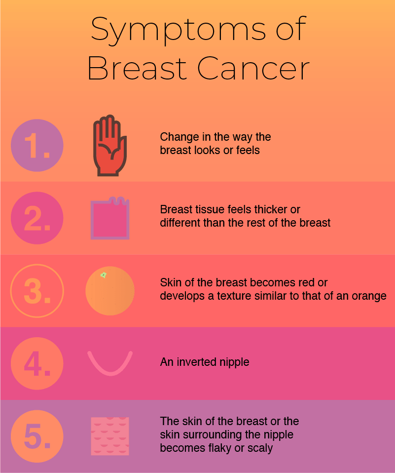
If you or your doctor finds evidence of a lump or tumor in your breast, the next step is to determine whether or not the lump is cancerous. Your doctor may choose one or more different ways to test for and diagnose breast cancer. At UVA Radiology, we know this can be a scary time of waiting. We want you to understand how the different types of tests work and what they involve so that you can go to your tests with confidence and assurance that your doctors are doing all they can to make an accurate diagnosis.
First Step: Finding A Tumor
Firstly, a breast lump or tumor must be found before a doctor will test the lump for breast cancer. If you’re a woman, checking for breast lumps should become a regular part of your life. Catching a tumor early can help prevent the spread of cancer and increase treatment.
The following are two of the most common ways that breast lumps are found, and should be performed regularly to monitor breast health.
Breast Exam
A breast exam can be performed by either you or a doctor. This exam involves feeling both your breasts and the lymph nodes under the arms to check for any abnormal growths or lumps.
Mammograms and Diagnostic Mammograms
Mammograms are x-rays of the breast, performed by radiologists. You should have a mammogram regularly to monitor your breast health. If a mass is detected in your breast, a doctor may order a diagnostic mammogram to test for cancer. Diagnostic mammograms are more in-depth than regular mammograms and take scans of the breast from multiple different angles, or a more zoomed-in scan of the breast where a lump is found.

Second Step: Testing for Breast Cancer
We know that finding a lump can be a frightening experience. If you find a lump, it’s important not to panic– most lumps turn out to be benign. Regardless, if you or a doctor finds evidence of a mass in your breast, further testing should be done to determine the nature of the tumor. There are a number of different options available to diagnose breast cancer, and you should be informed about the test your doctor has ordered so that you feel more prepared.
2) Breast Ultrasound

An ultrasound uses focused sound waves to create images of the inside of the body. Radiologists use ultrasound as a radiation-free way to determine the nature of a lump in the body–whether it is a tumor, a cyst, or something else entirely.
2) Breast MRI

MRI is short for Magnetic Resonance Imaging. When you have an MRI done, you’re put into a machine that creates images of the inside of your body for radiologists to interpret – in this case, your breast. Before having an MRI, patients are given an injection of dye to enhance the quality of the scan. A typical breast MRI takes about 30 minutes, but don’t worry about radiation. An MRI uses magnetic and radio waves to create an image instead of radiation, so you won’t be exposed to any radiation. Before you enter the MRI, you will be asked not have any metal on you – an MRI is basically a huge magnet!
3) Biopsy

A biopsy is a procedure where a doctor removes a piece of tissue and sends it to a lab for testing.
When taking a biopsy of a suspected tumor, your doctor will use X-ray to guide a specialized needle to take a sample of the lump itself. At this point, your radiologist will send the sample to a lab to be tested by a pathologist.
Biopsy is different from other tests because it yields a definitive result. It is more invasive, but the tests performed will show definitively whether or not the tissue is cancerous and can give valuable insight into how to treat it.
After Diagnosis
Once diagnosis has been completed, you and your doctor will discuss how to treat your condition. Treatment options depend on the type of breast cancer, how advanced the cancer is, your sensitivity to hormones, and your overall health. You have control of how you treat your breast cancer. Know what your options are before starting treatment and work with your doctor to figure out which course of action is right for you. To learn more about the getting screened for breast cancer at UVA, visit our website here.


