The term “nuclear” can sound rather intimidating as it often has negative associations with nuclear weapons. However, nuclear medicine is nothing to be intimidated by— it is not only safe, but it uses small amounts of radiation to do truly amazing things.
If you or a loved one has ever dealt with cancer, epilepsy, Parkinson’s disease, or any problems with organ function, they may have undergone a nuclear medicine procedure. At UVA Nuclear Imaging, nuclear medicine procedures are extremely helpful in determining organ structure and function, and are used to both diagnose and treat diseases.
What is Nuclear Medicine?
Nuclear medicine (Nuke for short) is a type of medical specialty that uses very small amounts of radiation administered inside your body to detect, diagnose, and even treat a multitude of diseases and conditions. Radiologists interpret the images from your nuke imaging exam to diagnose and treat your condition.

During a nuclear medicine procedure, doctors will use radioactive medicine in the form of injection, pill, or gas to target a part of your body and produce images that show your internal structures and their functions. These procedures can often detect diseases early, sometimes even before you experience any symptoms! Depending on why you need medical images, nuke may be the best option for imaging.
How Does It Work?
Nuclear medicine uses radiopharmaceuticals or radiotracers that carry a small dose of radiation. They are designed to travel to a particular part of your body so that they can gather the kinds of images the radiologist needs. Radiotracers can be injected into your bloodstream, swallowed in a pill, or inhaled as a gas.
Once the radiotracer is active in the part of your body you’re having imaged, a technologist will use a scanner and a computer to create the images of your organs and tissues. The scanners read the radiotracer to document the processes of organs and other functions in your body.

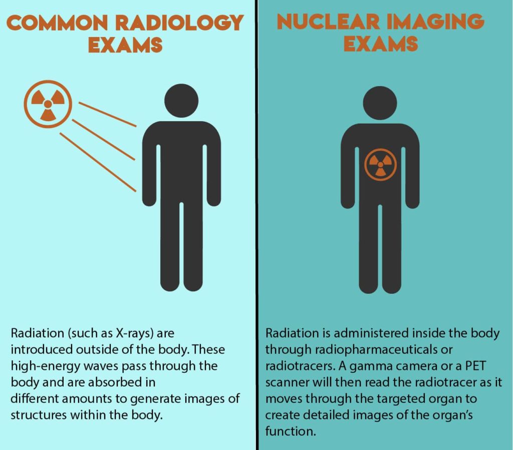
Your technologist could use a gamma camera, which looks like a CT machine (a giant circle with a doughnut hole that a table slides through), or a positron emission tomography (PET) scanner which also resembles a CT scanner. These machines do not use radiation waves themselves, but they can read the radiation in the radiotracer inside your body.
To reiterate, in nuclear medicine radioactive materials are introduced inside of the body. This differs from typical radiology imaging exams (such as X-rays or CTs) where radiation is introduced outside the body.

What Does Nuclear Medicine Look For?
Nuclear medicine procedures diagnose and treat diseases. They can produce images that radiologists can use to make a diagnosis based on structural appearance of your organs and how your organs, tissues, and bones are functioning. This is unlike other common imaging exams that cannot capture what your organs look like or how they’re actually working.
Nuke is used for detecting and staging cancer and other diseases, localizing tumors, monitoring cancer recurrences, and detecting some diseases in their earliest stages. Some of the things nuke can diagnose and treat:
- Epilepsy
- Parkinson’s disease
- Abnormal lesions
- Alzheimer’s disease
- Cancers
- Problems with transplanted organs
- Problems with heart, brain, and other organ function
Why and When is Nuclear Medicine Necessary?
Sometimes, nuclear medicine procedures are the best way to find and diagnose diseases in their earliest stages, while they are most treatable. Other times, they serve as a faster, non-invasive, less expensive alternative to exploratory surgery.

If your doctor is recommending you have a nuclear medicine procedure, it is probably the right choice for you. After all, these exams are the best at early disease detection and imaging the function of organs and tissues.
Is Nuclear Medicine Safe?
Nuclear medicine is in fact safe. Your doctors would not recommend something that would cause harm. There is not enough radiation in the radiotracers to hurt you, even over a long time; just enough radiation is used to get good enough pictures for an accurate diagnosis and no more.
At UVA Nuclear Imaging, we also control how much radiation is given to a particular patient by modifying the radiotracer to meet each patient’s individual needs. And the machines used to image your body do not use radiation, so you’re not getting a double dose. The radiation in the radiotracer is the only radiation you will receive in this exam. To learn more about nuclear imaging at UVA, click here.


