
Have you heard your doctor talk about or suggest a 3D mammogram, but aren’t sure how it differs from regular mammography? 3D mammography (also called Tomosynthesis) can help doctors detect breast disease earlier and more accurately than traditional mammography scans. At UVA Radiology, we want you to know about this state-of-the-art imaging technique and the advantages it offers.
What is Tomosynthesis?
Tomosynthesis (Tomo for short) is a big word for 3D mammography. Unlike traditional mammography, it takes multiple pictures of your breast tissue to create a three-dimensional image for your radiologist to interpret.
It can be used as a screening test (a regular test looking for breast cancer before symptoms are present) or a diagnostic exam (a test for diagnosing an issue). Tomo was developed to overcome some of the limitations of traditional 2D mammography and gives radiologists a clearer picture of your entire breast.
Tomosynthesis has been shown to:
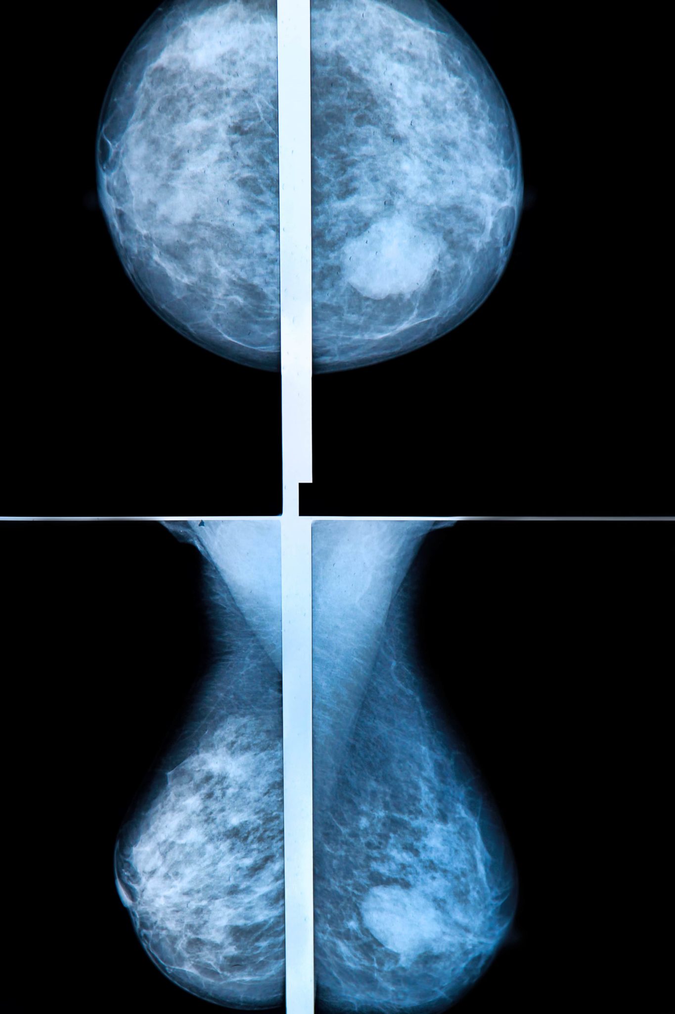
• Detect abnormalities that may have been hidden in dense breast tissue or folds due to compression during a traditional mammography
• Increase early detection of breast disease
• Decrease false positives, reducing unnecessary biopsies or additional tests
• Provide greater accuracy in determining the exact size, shape, and location of abnormalities
How Does It Work?
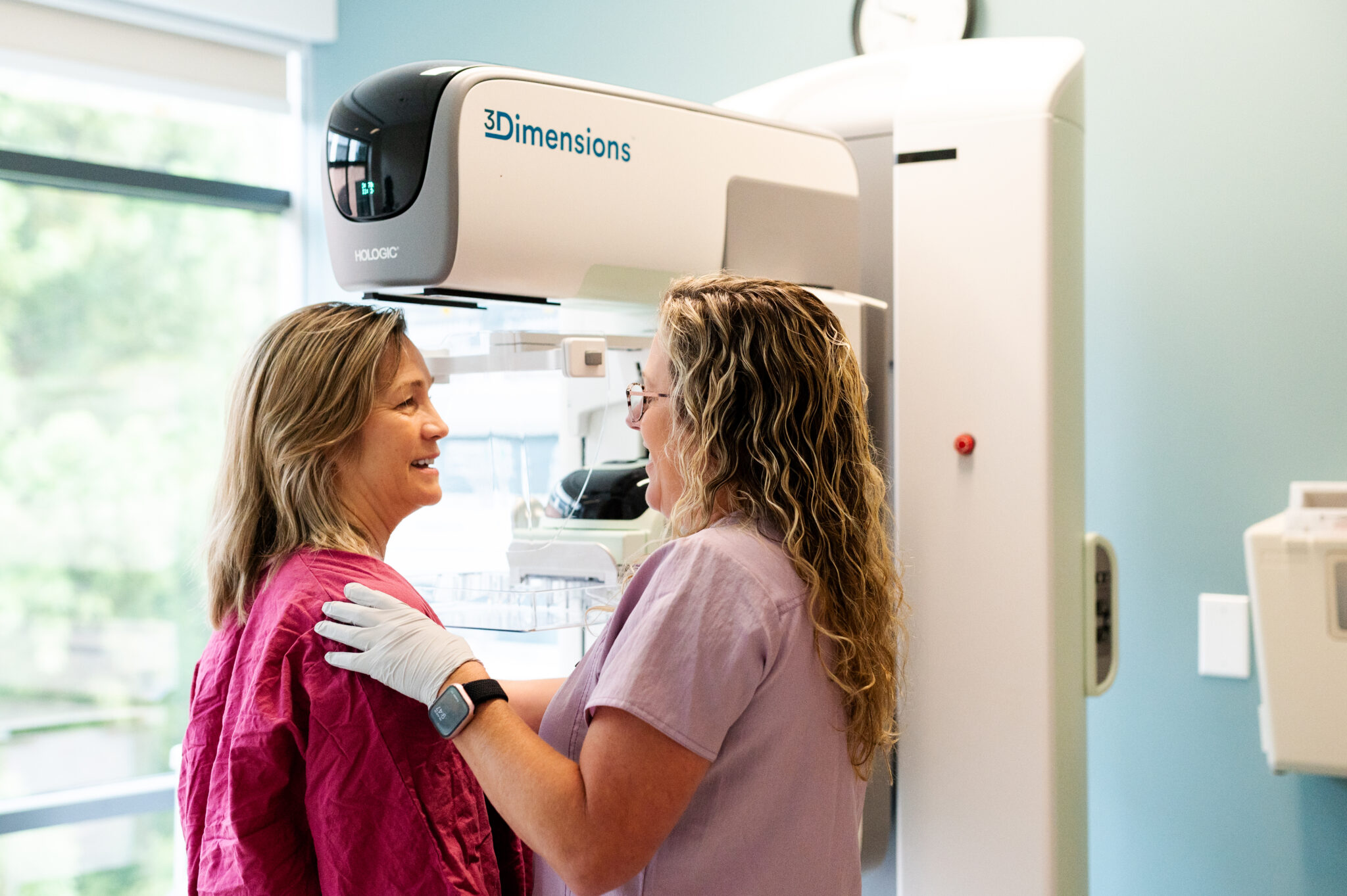
Tomosynthesis works very similarly to the way that traditional mammography works in terms of length of exam, machine, and your preparation. Both traditional mammography and Tomo require breast compression to obtain the correct images.
Tomo and traditional mammo differ in how the machine takes pictures. Traditional mammography compresses your breasts and uses low-dose x-ray to take four pictures: top, bottom, and each side. With tomosynthesis, a low-dose x-ray sweeps over the breasts in a tube and takes numerous pictures along the way. Then, a computer arranges the pictures into a 3D image set for the radiologist to read. Tomosynthesis can also produce the same two-dimensional pictures as traditional mammography.
Why Might I Need Tomo?
Women with dense breasts and those at high risk for breast cancer benefit most from Tomo. Early detection is the best defense against breast cancer, and breast cancer screening is the best way to detect breast cancer at its earliest stages when it is most treatable. Tomo can improve early detection and detection in dense breasts.
3 Advantages of Low-Dose 3D Mammography
1.) Less Radiation
Since the discovery of the X-ray researchers have been finding new ways to decrease radiation levels, as too much radiation can be potentially harmful to your health. When radiologists began using Tomo, patients were concerned due to the increased radiation exposure related to tomosynthesis.
At UVA Breast Care Center we use the latest technology, including our low-dose tomosynthesis which creates the same images as earlier tomosynthesis but with the radiation exposure level of traditional mammography. Since Low Dose Mammography requires only one exam, radiation exposure is cut significantly. “It’s about 40% less,” according to Breast Radiologist Dr. Brandi Nicholson in her interview with CBS19.
With Low Dose Mammography, you get all the benefits of a 3D mammogram (more accurate detection rates, fewer false positives, etc.) with radiation rates comparable to a regular 2D mammogram. It’s the best of both worlds!
2.) Less Time
With the latest technology, the exam takes only 3.7 seconds — just take a deep breath, count to four, and you’re done!
With a traditional 3D mammogram you receive a 3D scan then a 2D scan. Low Dose Mammography captures a 3D image and then generates a 2D image from the collected data, meaning your compression time decreases. Less time means less discomfort!
3.) Less Chance of Being Called Back for More Imaging
Low Dose Mammography has been proven to reduce false positive rates. This means that “we aren’t working up something that ultimately proves benign,” said Dr. Nicholson. The emotional roller coaster of the radiologist saying “I saw something” to later finding out “actually it was nothing” is something we all want to avoid.
Low Dose Mammography is also helping to increase cancer detection rates. The earlier cancer is found, the easier it is to treat. This is why yearly mammograms for women 40+ are so important!
Is the Cost of Tomo Covered?
Tomosynthesis is not yet the standard in breast imaging. Because of this, not all clinics offer it, and many insurance companies will not cover it. If your insurance company does not cover the cost, you can opt to pay an out of pocket fee of $82.00 to receive this type of imaging. If your doctor determines that Tomo is the best exam for you, the potential life-saving benefit is worth the cost.
Ready to make an appointment? We recommend annual mammography to women over age 40 or women who are at high-risk for breast cancer. Ask your doctor if Tomosynthesis is right for you. Call 434-924-1555 to make your appointment. For more information, visit the UVA Breast Care Program.